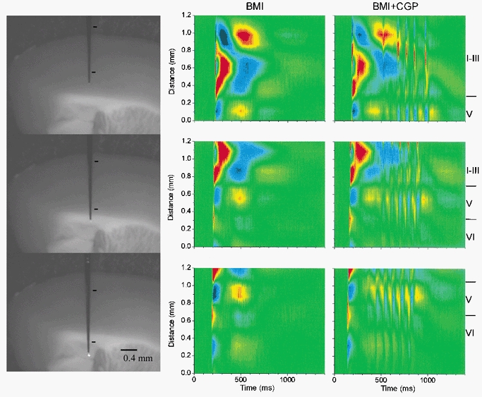
Figure 7.

CSD analysis of spontaneous discharges in agranular neocortex during BMI and during BMI+CGP35348
Left panels, photomicrographs of the slice preparation taken during the experiment showing the location of the 16-channel silicon probe used to derive the CSD analyses in the middle and right panels. The marks indicate the region corresponding to the CSD area. Middle panels, CSD analysis corresponding to spontaneous discharges generated in the agranular cortex during application of BMI. Each CSD analysis corresponds to a single spontaneous discharge recorded in the same cortical area; the only difference is the distance of the probe with respect to the pia mater. CSD analyses were derived from the spontaneous activity without averaging. Right panels, CSD analysis corresponding to spontaneous discharges generated in the agranular cortex during application of BMI+CGP35348, and recorded in the same locations as in the middle panels.
