Figure 2.

Effects of high perfusate flow rates
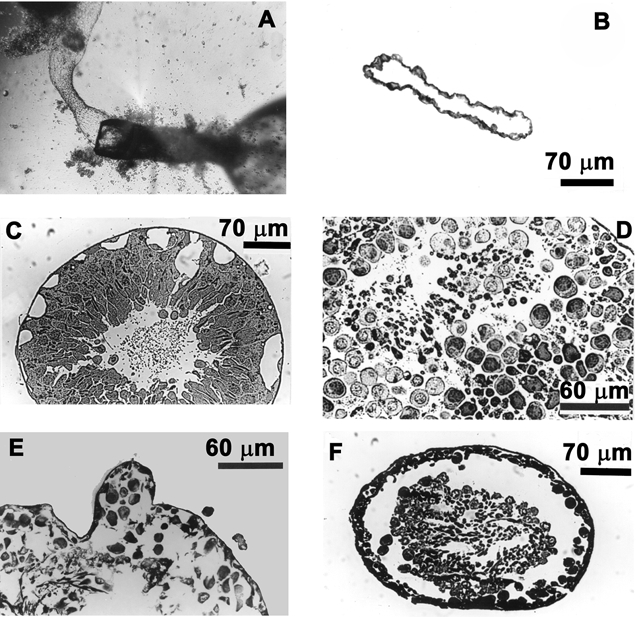
A, the perfusion pipette with the attached myoid tubule subsequent to the ejection of the germinal epithelium. B, cross-section of a seminiferous tubule exposed to high perfusate flow rates (0.68 μl min−1). Note that only the outer myoid layer remains resistant to high perfusate flow rates. C, decreasing the flow rate to 0.42 μl min−1 resulted in the formation of large vacuoles leading to the breakdown (D and E) and eventual ejection of the germinal epithelium (F).
