
Figure 3.
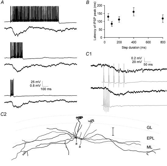
Time course of lateral IPSPs
A, lateral IPSPs were evoked in one mitral cell by trains of APs lasting 50–800 ms in a second cell. The time course and amplitude of the lateral IPSP (average of 7 sweeps) were unaffected by the duration of the train of presynaptic APs (single example sweeps shown). B, group data for four cells showing that in experiments similar to those shown in A the latency from the beginning of the presynaptic current injection to the peak of the lateral IPSP was unaffected by changing the duration of the presynaptic current step. C1, paired recordings from two mitral cells showing inhibitory coupling. Trains of 5 APs evoked at 20 Hz (top) or 100 Hz (bottom) resulted in IPSPs of similar amplitude and time course in the postsynaptic mitral cell. Postsynaptic traces are averages of 6 sweeps. C2, reconstructed morphology of the mitral cell pair from which the data in (C1) were recorded. Scale bar is 100 μm.