
Figure 1. Particles moving within a neurite of the cultured adult mouse dorsal root ganglion (DRG) cell, visualised by video-enhanced microscopy.
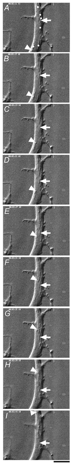
Serial images obtained at 4 s intervals indicate particles moving in anterograde (arrow) and retrograde (arrowhead) directions. Mitochondria (dots in A) were identified by their long-shape, large-size and saltatory movement. Scale bar, 2 μm.