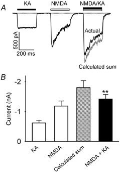
Figure 1. Kainate and NMDA-induced currents are sub-additive in hippocampal neurones.

A, current traces recorded from the same acutely isolated CA1 hippocampal neurone (VH = −60 mV) in response to rapid application of kainate (KA, 200 μM for 250 ms), NMDA (100 μM for 250 ms) and both agonists (NMDA + KA at the same concentrations for 250 ms). The grey trace is a calculated sum of the two individual current traces in response to kainate alone and NMDA alone. Note the difference between the current of the calculated sum (grey trace) and the actual current response to co-application of kainate and NMDA. To obtain full-sized NMDAR-mediated currents, the bath contained 20 μM glycine and zero magnesium. B, bar graph showing the average current amplitude induced by kainate alone (KA), NMDA alone (NMDA), the calculated sum (grey bar) and co-application of the two agonists (NMDA + KA, black bar). The current amplitude for co-application of NMDA and KA was significantly less than the calculated sum of the two currents induced by these two agonists individually (P < 0.01, n = 7).
