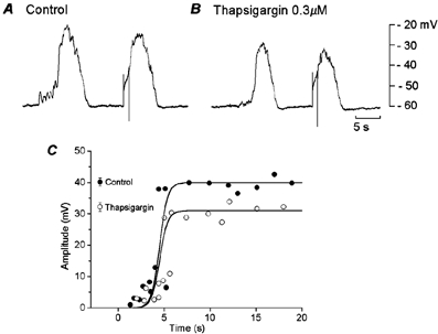
Figure 3. Effects of thapsigargin on evoked slow potentials.

During simultaneous recordings of membrane potential from two cells in a segment of circular smooth muscle tissue of the guinea-pig gastric antrum, muscle was stimulated by current pulses of 1 s duration and 3 nA intensity, in the absence (A, control) and presence of 0.3 μM thapsigargin (B). A and B were recorded from the same pair of cells. C, the relationship between the time of application of stimulating pulses after cessation of spontaneous slow potentials and amplitude of responses evoked by the pulses, in the absence (•, control) and presence of 0.3 μM thapsigargin (○).
