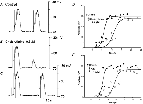
Figure 6. Effects of chelerythrine and bisindolylmaleimide I (BIM) on slow potentials.

A segment of circular smooth muscle tissue was stimulated by pulses (1.2 s duration, 2.5 nA intensity) at about 10 s (A and B) and 15 s (C) after the cessation of an evoked slow potential, in the absence (A, control) and presence of 0.3 μM chelerythrine (B and C). The amplitudes of the evoked responses were measured, in the absence (•, control) and presence (○) of 0.3 μM chelerythrine (D) or 0.2 μM BIM (E). Mean amplitudes (± s.d.) of the conditioning slow potentials are shown at the left hand sides of each graph (D, n = 9 for control and chelerythrine, P < 0.05; E, n = 9 for control and n = 12 for BIM, P < 0.05). A, B and C were recorded from the same pair of cells. D and E were obtained from different tissues.
