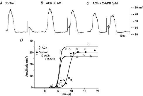
Figure 8. Effects of ACh and 2-APB on slow potentials.

An isolated segment of circular smooth muscle of guinea-pig gastric antrum was stimulated with depolarizing pulses (1.5 s duration, 2 nA) in the absence (A, control) and presence of 30 nm ACh (B) and ACh + 5 μM 2-APB (C). All responses were recorded from the same cell (the resting membrane potential, −64 mV). D, amplitudes of responses evoked by depolarizing pulses applied at various times after the cessation of a spontaneously generated slow potential, in the absence (•) and presence of ACh (○) and ACh + 2-APB (▵). The values shown at the left hand side of the graph indicate the amplitude of spontaneous slow potentials (mean ± s.d.; P < 0.05 compared to control).
