
Figure 1. Schematic diagram of the experimental layout.
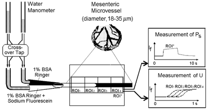
A single mesenteric microvessel (18–35 μm in diameter) was cannulated and perfused by a double-barrelled micropipette. One barrel was filled with the appropriate Ringer or Ringer-Locke solution while the other barrel contained the same solution to which sodium fluorescein had been added. Each barrel of the micropipette was connected through an electrically controlled cross-over tap to the water manometers. Pressures were set so that the vessel was perfused with solution from only one barrel of the pipette (nonfluorescent solution in the control state). When the cross-over tap was switched, the solution perfusing the vessel was immediately changed with minimal change in flow velocity. Images of the vessel and surrounding tissue were observed through a stereomicroscope and acquired by CCD camera to be analysed off-line. The time course profile of the fluorescent light intensity, If, for a region of interest (ROI*) was used to evaluate both PS and U.