
Figure 6. Synaptic protection in Wlds mice depends on synaptic maturity and not the age of the animal.
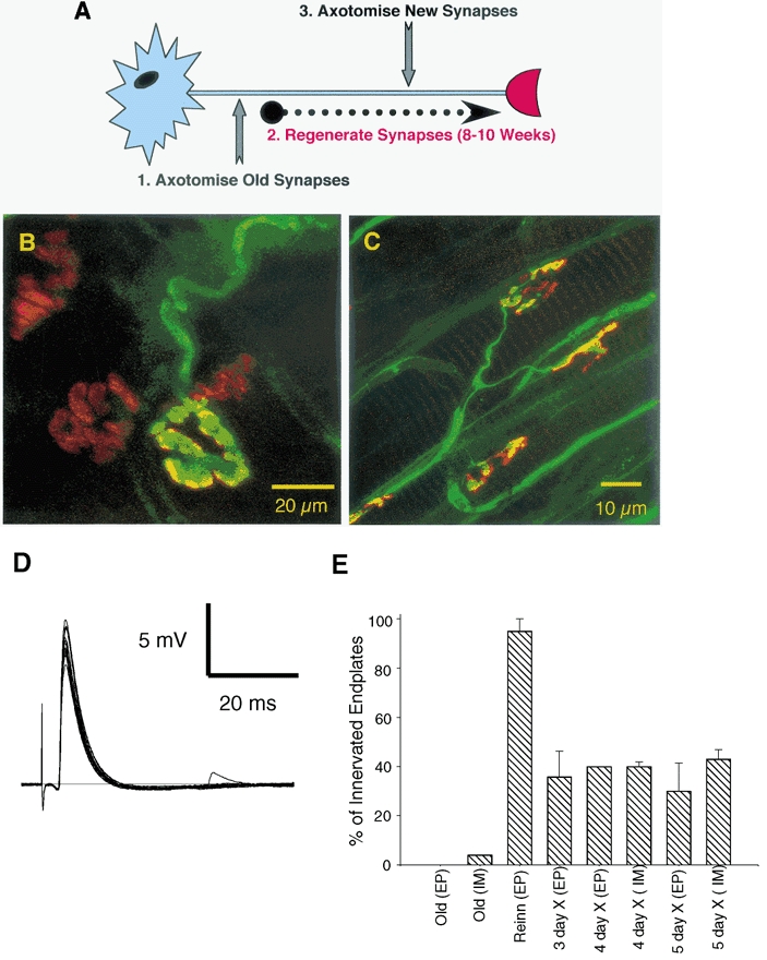
A, schematic representation of the experimental protocol used to assess the role of synaptic maturity versus chronological age on nerve terminal preservation following axotomy. The Wlds mice used were > 7 months old, therefore permitting a test of the responses to axotomy at immature (i.e. 2–4-week-old), regenerated synapses, in mature mice. B, confocal micrograph of an immunocytochemically labelled 7 month Wlds FDB muscle, 3 days post axotomy. One of the few remaining terminals from this preparation is shown, with a characteristic bloated appearance, surrounded by three vacated endplates. C, confocal micrograph of a regenerated synapse from a 14 month Wlds FDB muscle, 5 days post axotomy. Axons and motor nerve terminals were labelled immunocytochemically (NF and SV2; FITC) and acetylcholine receptors were labelled with TRITC-conjugated α-BTX. Note that all endplates shown are occupied and that the uppermost instance shows evidence of partial occupancy. D, electrophysiological recording from a 14 month Wlds FDB muscle fibre with regenerated synaptic connections, 5 days post axotomy, showing robust synaptic transmission. E, graph showing the percentage of fibres with synaptic innervation, as assessed by electrophysiological (EP) and immunocytochemical (IM) techniques, in old (non-regenerated) Wlds FDB muscle fibres at 3 days post axotomy; in reinnervated muscle fibres 8 weeks after the first lesion but before a second lesion; and in reinnervated endplates at 3–5 days after the second lesion. Note the rapid and almost complete loss of innervation following axotomy at the axotomised ‘old’ synapses (compare with Figure 4) and the retention of > 30 % of regenerated synaptic connections for up to 5 days post axotomy.