
Figure 7. Age-dependent synaptic protection in Wld transgenic mice.
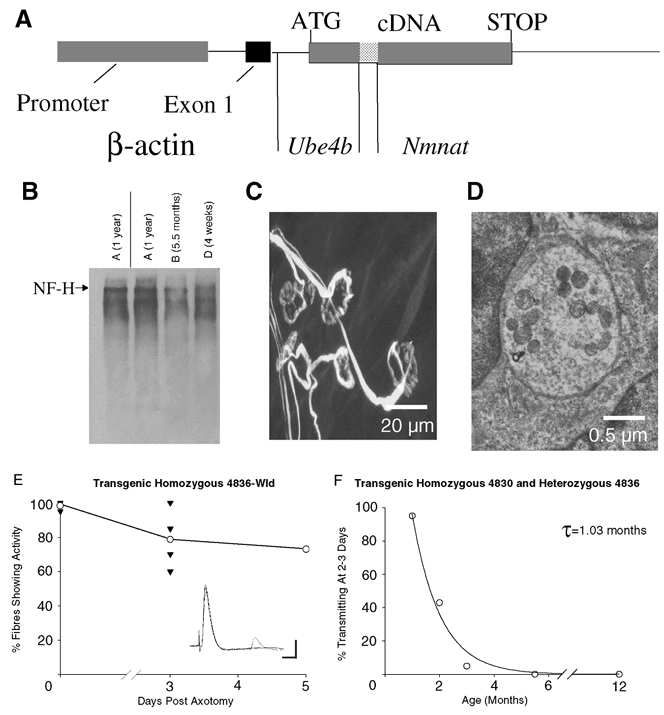
A, the transgene construct for Ube4b/Nmnat (Wld) transgenic mice. The chimeric cDNA was expressed with non-coding exon 1 of β-actin under the control of a human β-actin promoter and terminated with the SV40 polyadenylation signal. B, Western blot showing age-independent preservation of heavy chain neurofilament proteins (NF-H) in the distal stump of 4836 Wld transgenic mice sciatic nerve, 3 days post axotomy. C, confocal micrograph of immunocytochemically labelled (NF, SV2 and α-BTX) persistent synapses, 5 days post axotomy in a 4836 Wld transgenic lumbrical muscle. All of the endplates shown are either fully occupied or are missing only a small proportion of their nerve terminal. Examples of endplates with less than 50 % occupancy as well as evidence for retraction bulb formation were also found (data not shown). D, electron micrograph of a retained synaptic bouton from a 4836 homozygous Wld transgenic mouse FDB muscle, 5 days post axotomy, showing intact membranes and synaptic vesicles. E, time course of the loss of innervation following axotomy in 4836 Wld transgenic mice. The inset illustrates an electrophysiological recording showing robust synaptic transmission from a 2-month-old 4836 Wld transgenic FDB muscle fibre, 5 days post axotomy. Scale bars = 5 mV (vertical); 10 ms (horizontal). F, age dependence of synapse loss in 4830 and 4836 heterozygous Wld transgenic mice. The incidence of fibres showing synaptic responses 2–3 days after axotomy declined exponentially with age with a time constant (τ) of ∼30 days, similar to the natural mutant (compare with Fig. 3).