
Figure 8. Persistence of neuromuscular junctions following axotomy in thy1-CFP/Wld-homozygous mice.
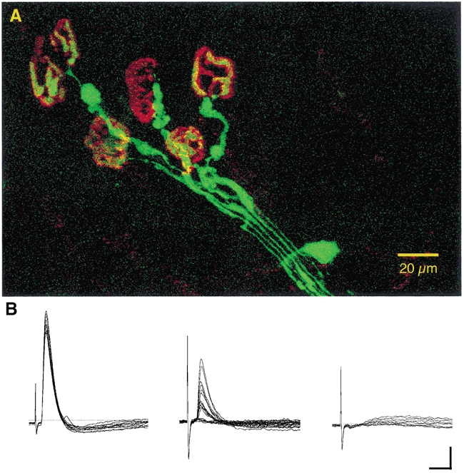
A, confocal projection image of endogenous CFP fluorescence of a group of axons and terminals supplying motor endplates, counterstained with TRITC-α-bungarotoxin, in an isolated, unfixed lumbrical muscle, 4 days after axotomy. CFP fluorescence has been pseudo-coloured green, TRITC fluorescence red. B, electrophysiological recordings of robust (left) and weak (middle: low quantal content, failures) synaptic responses recorded from FDB muscle fibres in the same axotomised foot. The record on the right shows that, as in normal Wld mice, a few fibres fail to respond, corresponding to unoccupied endplates. Scale bars = 5μm (vertical); 10 ms (horizontal).