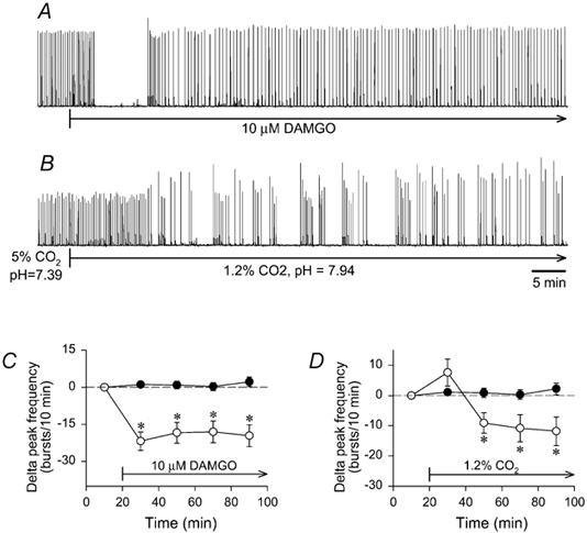
Figure 7. Strychnine-bicuculline rhythm altered by μ-opioid receptor activation and low PCO2.

Brainstems were exposed to bicuculline and strychnine (50 μM each) for 1.5-2.0 h to establish a stable bicuculline-strychnine rhythm before DAMGO or low PCO2 application. A and B, traces of integrated hypoglossal nerve activity produced during synaptic inhibition blockade by individual brainstems are shown. A, DAMGO (10 μM) application abolished rhythmic activity for 8.4 min, and decreased frequency by 19 ± 4 bursts (10 min)−1 for the next 60 min. B, switching from standard solution containing bicuculline and strychnine (5 % CO2; pH 7.39) to a similar solution bubbled with 1.2 % CO2 (pH 7.94) decreased frequency by 33.7 ± 1.2 bursts (10 min)−1 within 10 min. For both C and D, the frequency of the steady-state bicuculline-strychnine rhythm in control brainstems (•; n= 12) was 17 ± 2 bursts (10 min)−1 at the zero time point, and remained within 17.4-18.5 bursts (10 min)−1 for the next 80 min. All data were averaged in 20 min bins. C, continuous DAMGO application (10 μM, 80 min) reduced the steady-state bicuculline- strychnine frequency of 22.1 ± 2.4 bursts (10 min)−1 by 19.4 ± 0.8 bursts (10 min)−1 during 80 min application (○; n= 8). D, switching from 5 % to 1.2 % CO2 (80 min) increased the steady-state bicuculline- strychnine frequency of 21.5 ± 1.8 bursts (10 min)−1 at the zero time point by 7.6 ± 4.5 bursts (10 min)−1 during the first 20 min before causing a decrease of 10.5 ± 0.8 bursts (10 min)−1 during the next 60 min (○; n= 8). *P < 0.05 compared to the first 20 min for delta peak frequencies.
