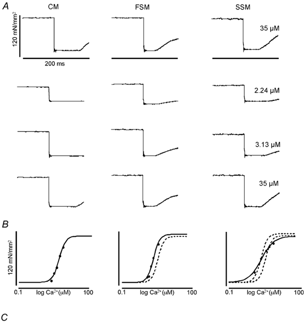
Figure 1. Method used to measure active force development under maximal and submaximal activation in rat cardiac trabeculae (CM), rat psoas (FSM), and soleus (SSM) muscle fibres.

A, original recordings of force, normalized as force (unit area−1, at the indicated [Ca2+] (in μM) to the right of the tracings for each striated muscle type (SL = 2.10 μm). A quick release was used to determine zero force level (see Methods). The quick release step size depended on muscle type and [Ca2+]; it was 10–20 % of muscle length. Calibrations as indicated. B, Ca2+-force relationship, constructed from data obtained as illustrated in A. The continuous lines indicate the Hill fit to the data obtained from the indicated muscle type (above each data column); the dotted lines represent the Hill fit obtained from CM (middle panel), or CM and FSM (right panel). C, typical CCD images of X-ray diffraction patterns obtained from each muscle type at the same SL (SL = 2.10 μm) indicative of the change in lateral separation of the myofilaments in each muscle. The 1,0 and 1,1 equatorial reflections are as indicated.
