
Figure 1. Different effects of hypoxia on isolated NTS neurones in a brainstem slice.
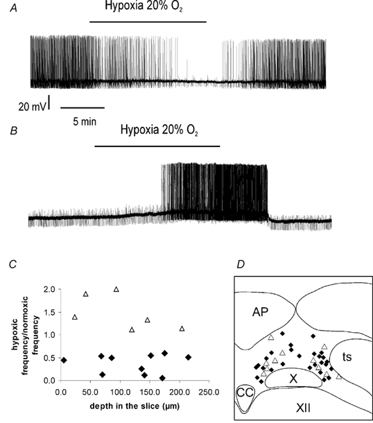
A, example of a neurone inhibited by hypoxia (intracellular recording). Moderate hypoxia (20 % O2) elicited a reversible hyperpolarisation accompanied by a reduction of spontaneous activity and of input resistance. B, example of a neurone excited by hypoxia (intracellular recording). Moderate hypoxia elicited a reversible depolarisation accompanied by an increase of spontaneous activity. C, type of neuronal responses to hypoxia, excitation or inhibition, according to the depth of the recording in the slice, expressed as the firing frequency ratio between normoxia and hypoxia (extracellular recording, n = 16). ♦, neurones inhibited by hypoxia; ▵, neurones excited by hypoxia. D, localisation of recorded neurones in the NTS (n = 38). ♦, neurones inhibited by hypoxia; ▵, neurones excited by hypoxia. AP, area postrema; CC, the medullary central canal; ts, the tractus solitarius; X, the dorsal motor vagal nucleus; XII, the hypoglossal nucleus.