
Figure 3. Intracellular pH in oocytes as a function of extracellular pH and butyrate.
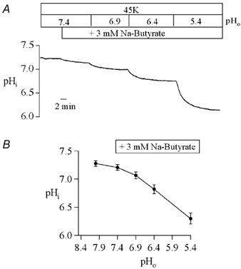
A, typical titration in a Xenopus oocyte injected with wild-type Kir1.1a cRNA. Identical results were obtained for oocytes injected with Kir1.1a 351X cRNA. Butytrate was added and the pH of the 45 mm K+ bathing solution was changed as indicated. Intracellular pH was measured with intracellular pH-sensitive microelectrodes every 2 s for the duration of the record. B, the mean steady-state pHi in each of the bathing solutions is summarized.