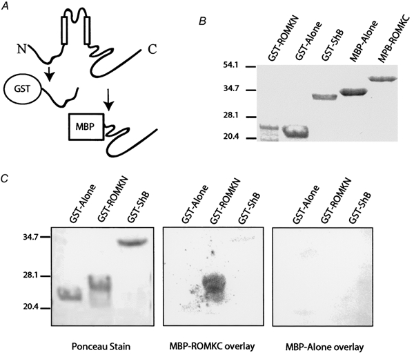
Figure 7. Cytoplasmic NH2- and COOH-terminal domains of Kir1.1 interact in vitro.

A, cartoon illustrating the cytoplasmic domains of Kir1.1 and the corresponding bacterial fusion proteins. The NH2-domain was made as a GST fusion and the COOH-domain was made as a MBP fusion. B, purified recombinant domains resolved by SDS-PAGE and visualized by Coomassie Brillant Blue staining. C, gel overlay assay: GST alone or GST fusions of the Shaker B cytoplasmic NH2-terminus (GST-ShB) or the Kir1.1 N-terminus (GST-ROMKN) were resolved by SDS-PAGE, transferred to nitrocellulose, renatured, visualized by Ponceau staining (left) and then blotted with either the MBP protein alone (right) or MBP fusion of the Kir1.1 COOH-terminal domain (MBP-ROMKC, middle). After extensive washes, bound MBP protein was detected using an anti-MBP antibody.
