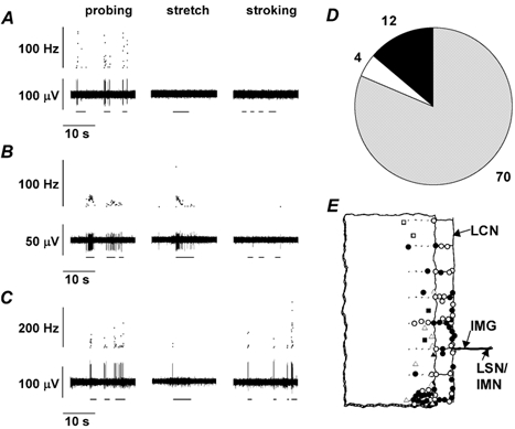
Figure 1. Three types of fibre were classified on the basis of their responses to mechanical stimuli.

In A, B, and C upper traces show instantaneous frequency and lower traces show raw electrophysiological data. A, a serosal receptor is activated only by probing the tissue (horizontal bars). B, a muscular receptor is activated by probing and by a maintained circular stretch of 8 mm; the response adapts rapidly during the stimulus (horizontal bars). No increase above baseline activity occurred in response to mucosal stroking. C, a mucosal receptor responds to probing and to stroking of its receptive field with a von Frey hair giving a force of 0.1 mN. D, the majority of fibres encountered were serosal receptors (grey segment; numbers shown around pie chart), the remainder being split between muscular (open segment) and mucosal receptors (filled segment). E, the distribution of receptive fields was concentrated near the mesenteric border and on the mesentery itself. Circles, serosal afferents; squares, muscular afferents; triangles, mucosal afferents; filled symbols indicate responsive to 5-HT agonists, open symbols indicate unresponsive. LSN/IMN, lumbar splanchnic nerve/intermesenteric nerve; IMG, inferior mesenteric ganglion; LCN, lumbar colonic nerves.
