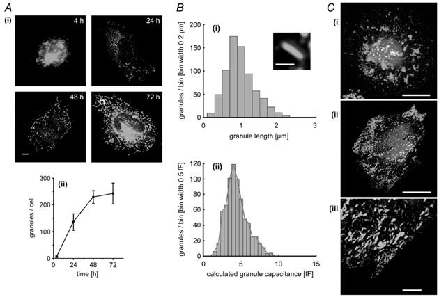
Figure 2. Time-dependent accumulation of Weibel-Palade bodies (WPb), distribution of WPb Cm, and subcellular localisation of von Willebrand factor (vWf) and tissue plasminogen activator (t-PA).

A(i), representative fluorescence images of fixed HUVEC that have been stained with a specific antibody to vWf at the times indicated. Images are projections of three to five, 0.2 μm optical sections, the scale bar indicates 5 μm. The mean number of vWf-immunopositive granules (WPb) counted in 12 cells at each of the indicated times is plotted in (ii) (means ± s.d.). B(i), the distribution of lengths of WPb measured from images such as those shown in (i) with an example (inset) of an average WPb from the 72 h cell in A(i) (indicated by the white box). The scale bar is 1 μm. The calculated distribution of WPb Cm derived from the data in B(i), assuming a specific Cm of 8 fF μm−2 is shown in (ii). A red line is drawn through the peak of each bin. C(i) and (ii), HUVEC at 4 and 24 h, respectively, labelled with specific antibodies to both t-PA (red) and vWf (green). An expanded region of the image in (ii; - 24 h cell) shows no co-localisation of t-PA and vWf. Images are projections of three, 0.3 μm optical sections, the scale bar being 20 μm in (i) and (ii) and 5 μm in (iii). Nuclear DNA was labelled with Hœchst 33342 (blue).
