
Figure 8. Brief elevation of [Ca2+]i to high levels does not evoke WPb exocytosis.
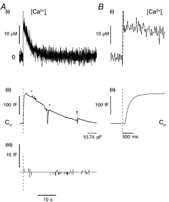
A, a continuous record of [Ca2+]i (i) and Cm (ii) from a cell that had been 72 h in culture, voltage clamped at -50 mV and dialysed with a solution containing 500 μm furaptra and calcium-DM-nitrophen (2.4:4 mm). A single, brief (1 ms) pulse of near-UV light (1500 μF, 200 V) was applied at the time indicated by the vertical dashed lines on the records in A(ii) and (iii) and B(i) and (ii). The detected Cm steps (exocytotic represented by upward lines, and endocytotic by downward lines) are shown in (iii). The area between the stars was excluded from step analysis due to a series of large Im fluctuations that might have produced spurious changes in Cm. B, the early part of the [Ca2+]i (i) and Cm (ii) response on an expanded time scale. The p-p noise was 0.88 ± 0.30 fF for the period shown. †, the point at which a CT and Rs compensation was applied.