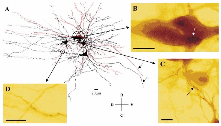
Figure 2. Putative sites of coupling between motoneurones.

A, camera lucida reconstruction of dye-coupled motoneurones from a P8 MK801-treated preparation. The impaled motoneurone is shown in red and a cluster of four dye-coupled cells are shown in black. Note the extensive overlap of the dendritic trees of the motoneurones containing several sites of close apposition as illustrated at the light microscope level (B-D). B, somato-somatic and somato-dendritic (white arrow) sites of close apposition. C, proximal dendro-dendritic sites of close apposition (arrow). D, distal dendro-dendritic site of close apposition. Note that the axons of two of the motoneurones are seen leaving the spinal cord (arrows). Scale bars in B, C and D: 20 μm. R, rostral; C, caudal; D, dorsal; and V, ventral.
