
Figure 1. Induction of Ca2+ alternans.
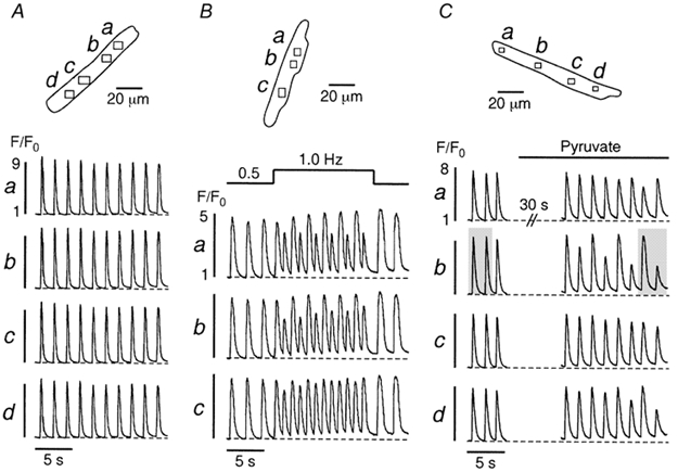
A, under control conditions (glucose-containing Tyrode solution) various regions (a-d) of an atrial myocyte exhibit uniform [Ca2+]i transients. An increase in stimulation frequency from 0.5 to 1.0 Hz (B) or application of pyruvate-containing (10 mm) solution (C) elicit Ca2+ alternans. Stimulation frequency in A and C, 0.6 Hz. The shaded areas in C mark the [Ca2+]i transients shown in more detail in Fig. 2.