
Figure 5. Neighbouring regions within an atrial myocyte alternate out-of-phase.
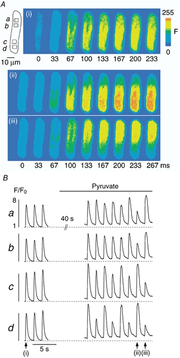
Ca2+ alternans was induced by application of pyruvate. A, series of fluo-4 fluorescence images recorded under control conditions and during Ca2+ alternans. The images illustrate the rising phase of the [Ca2+]i transients marked by the arrows in B. B, subcellular [Ca2+]i transients recorded from the regions marked by the rectangles a-d (A). [Ca2+]i images and subcellular [Ca2+]i transients reveal that the time of onset, the magnitude, and the phase of Ca2+ alternans exhibit large subcellular variations and that the upper and the lower half of the cell alternate out-of-phase. Stimulation frequency, 0.6 Hz.