
Figure 7. Subcellular Ca2+ alternans causes intracellular propagating Ca2+ waves.
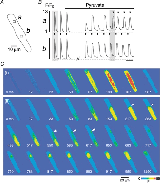
An atrial myocyte (A) is challenged with pyruvate (10 mm). B, subcellular [Ca2+]i transients recorded from the regions a and b marked by the rectangles in A. C, cellular fluo-4 fluorescence images recorded under control conditions (i) and during pyruvate-induced Ca2+ alternans (ii) (shaded regions marked in B). 0 ms refers to the image recorded immediately preceding the first detectable increase of [Ca2+]i. In the presence of pyruvate region a exhibits Ca2+ alternans, whereas region b does not (B). This pattern leads to a steep [Ca2+]i gradient between the two regions (C, panel (ii), arrows at 217 and 283 ms). A delayed localized [Ca2+]i increase at the border zone (C, arrowheads at 550, 583 and 617 ms) gives rise to a propagating Ca2+ wave into the upper part of the cell (C, 650–1250 ms). In panel Ba electrical stimulations are indicated by filled circles and delayed Ca2+ waves are marked by asterisks. Stimulation frequency, 0.6 Hz.