
Figure 3. Action potentials and current signatures in cold-sensitive and cold-insensitive neurones.
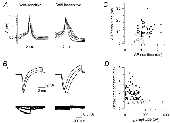
A, action potentials in 10 randomly selected cold-sensitive neurones (left) and 10 cold-insensitive neurones (right) in response to 5 ms depolarising stimuli, aligned so that the peak occurs at the same time. Resting potentials were between −40 and −70 mV; in some cases the beginning of the stimulus is beyond the left edge of the trace. Bi, transient inward currents in one cold-sensitive (left) and one cold-insensitive neurone (right); from a holding potential of −60 mV, after a 150 ms prepulse to −80 mV, the potential was stepped to −10, 0 and +10 mV for 6 ms. The decay time constant was measured for the most positive pulse. Bii, hyperpolarisation-activated current Ih in one cold-sensitive (left) and one cold-insensitive neurone (right); from a holding potential of −60 mV, the potential was stepped to −70, −90 and −110 mV for 500 ms. Current amplitude was measured for the most negative pulse. Whole-cell (not perforated-patch) recordings, K2SO4 pipette solution; recordings were corrected offline for leak and capacitive transients, and any residual capacitive transients are blanked. C, relation between action potential rise time and the amplitude of the after-hyperpolarisation (AHP) in cold-sensitive (○) and cold-insensitive (•) neurones. D, relation between Ih amplitude and decay time constant of the transient inward current (see B) in cold-insensitive neurones (•) and cold-sensitive neurones sensitive (□) and insensitive (▵) to capsaicin.