
Figure 7. Effect of isoproterenol upon voltage-dependent inactivation of ICa,L.
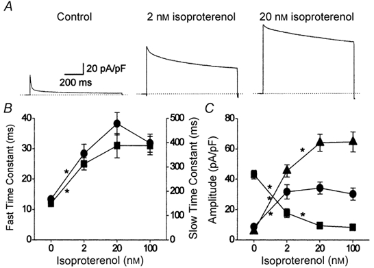
Cells were bathed in zero calcium extracellular solution and currents were evoked by voltage-clamp steps to +80 mV (A). The decay of the evoked cell currents was fitted by a bi-exponential function (Origin 4.1) and the components of that function were extracted (B and C). A, representative cell current records obtained from three different myocytes exposed to either basal conditions in the absence of any external agonist (Control; left), 2 nm isoproterenol (centre) or 20 nm isoproterenol (right). The dotted lines indicate the 0 pA current level and the time and current scales shown on the left apply to all three traces. B, effect of isoproterenol upon the fast (left axis; ▪) and slow (right axis; •) time constants of decay. Note the different y-axis scales for the two time constants. C, effect of isoproterenol upon the initial amplitude of fast (▪) and slow (•) time-dependent, and the time-independent (▴), components of decay. Control (0 nm isoproterenol), n = 8; 2 nm isoproterenol, n = 7; 20 nm isoproterenol, n = 6; 100 nm isoproterenol, n = 7. * P < 0.05, for comparison between adjacent groups with one-way ANOVA (Origin 4.1).