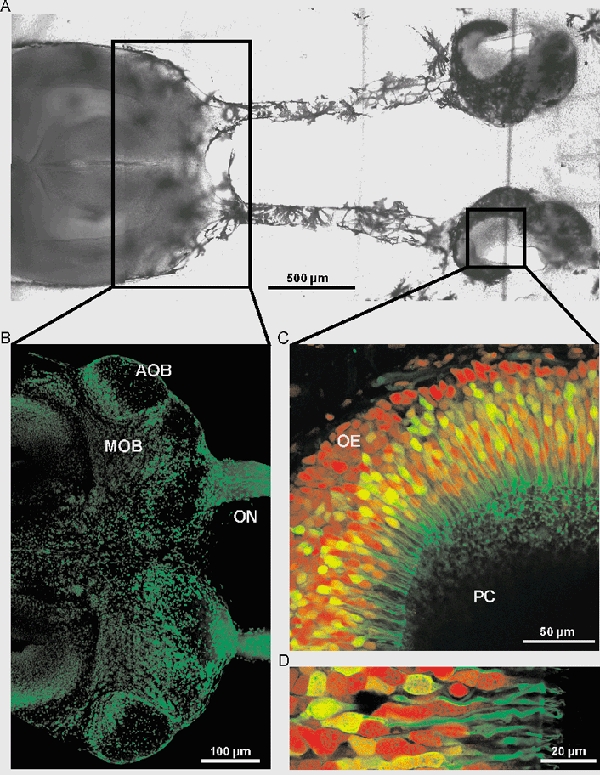
Figure 1. Slice of the olfactory mucosa and the olfactory bulb.

A, image of the slice of the olfactory mucosa and the olfactory bulb. B, slice of the anterior part of the brain including the olfactory nerve (ON), the main olfactory bulb (MOB) and the accessory olfactory bulb (AOB) stained with propidium iodide. C, horizontal overview of the olfactory epithelium (PC, principal cavity and OE, olfactory epithelium). The neurons were backfilled through the nerve using biocytin-avidin staining (green fluorescence), and the slice was counterstained with propidium iodide (red fluorescence). D, higher magnification of C, showing ORNs with dendrites, dendritic knobs and cilia.
