
Figure 7. Conductance of the leak, gleak, as a function of time associated with single-cell defects.
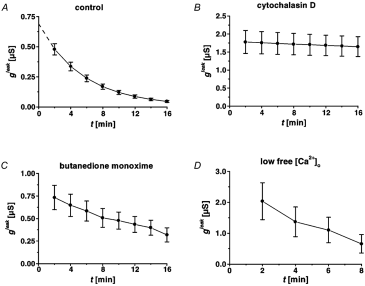
Time course of restitution in HT-29/B6 cells (28th-31st passage), under control conditions (A), after incubation with cytochalasin D (B), with 2,3-butanedione monoxime (C), and in low free [Ca2+]o Ringer solution (D). Under control conditions (A), repair resulted in exponential decline of gleak, which allowed extrapolation (dashed line) of the initial (t + 0) leak at 0.69 ± 0.06 μS. Sealing of the leak (described by gleak) was blocked after inhibition of actin polymerization (B), and was slower after impairment of myosin ATPases (C), but proceeded at the same speed in low Ca2+ Ringer solution (D).