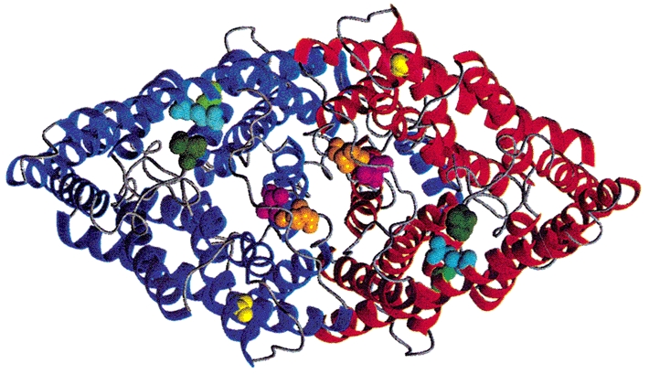
Figure 7. Location of residues at which mutations cause hyperpolarisation-induced activation.

Backbone fold of the Salmonella typhimurium ClC dimer, StClC (Dutzler et al. 2002), shown from the extracellular side. The locations of corresponding hClC-1 mutations are shown as space-filling models: A52 (S132C), light green; E54 (D136G), cyan; R147 (K231C), dark green; G372 (G499R), yellow; T416 (T550M), magenta; N418 (Q552R), orange. This figure was generated with the program MOLMOL (Koradi et al. 1996).
