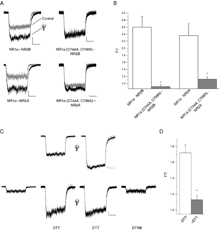
Figure 1. The redox site on the NR1 subunit of the NMDA receptor is required for light sensitivity.

A, whole-cell NMDA (10 μm)-induced currents from receptors NMDA receptors transiently expressed in CHO cells (membrane potential: −60 mV in this and all subsequent figures). Sample traces represent currents before (grey trace) and shortly after (5 s; black trace) a brief light pulse (2 s, > 280 nm). Similar effects were seen in 5-12 cells for each subunit configuration. Scale bars: 100 pA and 1 s. B, peak response amplitudes for responses such as those seen in A were measured and a post-flash to pre-flash (I*/I) ratio was calculated (* P < 0.05, significantly different from wild-type control, Student's two tailed t test; n = 5–12 cells). C, top, NMDA-elicited responses from NR1a-NR2B receptors both before and after a brief flash of light (light bulb; 2 s, > 280 nm) Scale bars: 500 pA and 500 ms. C, bottom, NMDA-mediated responses from NR1a-NR2B receptors before and after 8 min of 5 mm DTT incubation. Chemically reduced receptors were then exposed to brief flash (2 s, > 280 nm) followed by chemical oxidation with 500 μm DTNB (2 min). Scale bars: 100 pA and 1 s. D, I*/I ratios demonstrating a significant decrease in light sensitivity following chemical reduction (* P < 0.05, significantly different from ‘-DTT’ control, Student's two tailed t test, n = 5–7 cells).
