
Figure 2. Lobuloalveolar morphology and XOR localization in alveolar epithelial cells from pregnant and lactating mice.
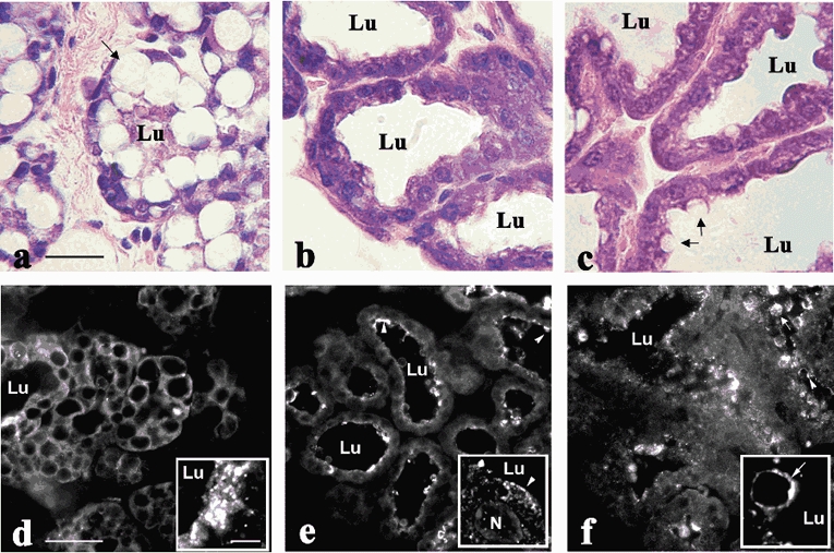
Haematoxylin/eosin staining (a-c; scale bar + 25 μm) of mammary gland sections from mice at day 19 of pregnancy (a), day 1 of lactation (b) and day 10 of lactation (c). The presence of large intracellular lipid droplets (arrow, a), the small lumen (Lu) size and the lack of luminal material indicate the absence of significant secretory activity at this period. The significant increase in luminal size and the loss of large intracellular lipid droplets in glands at day 1 of lactation indicate active secretion (b). At day 10 of lactation, lipid droplets in the process of being secreted can be seen at the apical membrane (arrows, c). Immunolocalization of XOR (d-f; low power images, scale bar + 50 μm) in perfusion-fixed mammary glands from mice at day 19 of pregnancy (d), day 1 of lactation (e) and day 10 of lactation (f). Insets in d-f show higher power images of XOR localization (scale bar + 1 μm). Arrowheads indicate apical membrane association of XOR, arrows show XOR labelling of secreted lipid droplets. The arrow in the inset in panel f indicates XOR on the surface of a lipid droplet in the process of being secreted.