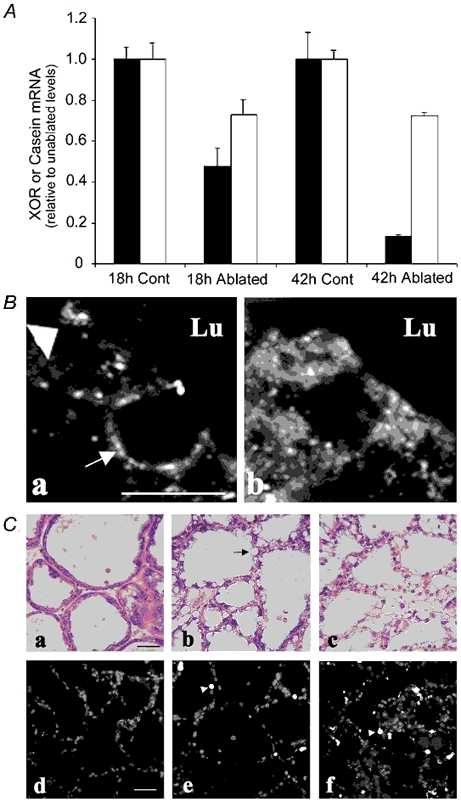
Figure 4. Effects of milk stasis on XOR expression and localization.

A, relative steady-state mRNA levels of XOR (▪) and β-casein (□) in mammary glands from paired unablated (control) and nipple-ablated (ablated) animals at 18 and 42 h after nipple ablation on day 15 of lactation. RNA levels determined by RT-PCR are shown as the percentage of the respective control values at each time point. B, immunolocalization of XOR in alveolar epithelial cells (a and b, scale bar + 2 μm) from unablated mammary glands (a) and nipple-ablated mammary glands 18 h after ablation on day 15 of lactation (b). The large arrowhead in a indicates XOR at the apical membrane, and the arrow in a shows XOR staining around a lipid droplet in the process of being secreted into the lumen. Note the absence of XOR at the apical membrane in ablated tissue. C, histological and terminal deoxynucleotidyl transferase-mediated deoxyuridine triphosphate nick-end labelling (Tunel) analysis of mammary tissue after nipple ablation. Panels a-c show haematoxylin/eosin staining of mammary glands from unablated animals (a), animals nipple ablated for 24 h (b) and 48 h (c). Scale bar + 20 μm. Panels d-e show Tunel staining in similar sections from unablated glands (d) and glands 24 h (e) and 48 h (f) after nipple ablation. The arrowheads indicate Tunel-positive nuclei. Scale bar + 40 μm.
