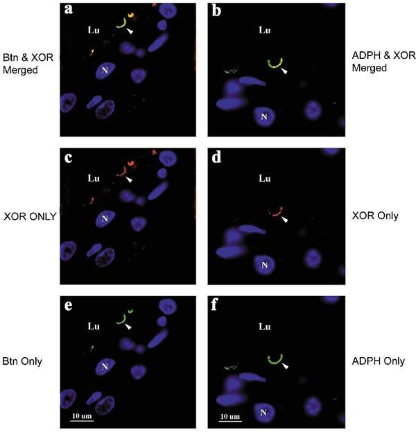
Figure 5. Colocalization of XOR, butyrophilin (Btn) and adipophilin (ADPH) in lactating mouse mammary tissue.

XOR was separately immunolocalized with Btn or ADPH in paraffin-embedded mammary tissue from mice at day 10 of lactation. XOR immunostaining was detected with cy3-labelled secondary antibodies (red), and Btn and ADPH immunostaining were detected with fluorescein isothiocyanate (FITC; green)-labelled secondary antibodies, as described in Methods. a, merged image of XOR and Btn staining; c, XOR only staining; e, Btn only staining; b, merged image of XOR and ADPH staining; d, XOR only staining; f, ADPH only staining. Nuclei (N) and luminal regions (Lu) are indicated. Scale bar + 10 μm.
