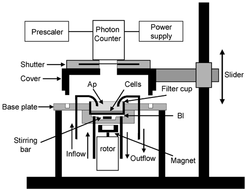
Figure 1. Schematic diagram of the experimental setup for measuring luminescence.

The entire filter cup that supports the epithelium is mounted in a holder that separates the apical (Ap) and basolateral (Bl) compartments. Perfusions of each chamber half proceed through black light-tight tubing. The volume of the basolateral compartment was 1 ml. The diffusion of ATP into the basolateral bath through the Anopore filter was accelerated by vigorously mixing the solution with a magnetic stirring bar coupled to a rotating magnet. The volume of the solution in contact with the apical side of the epithelium was 2 ml. The photon counter tube protected by a shutter device was mounted on a support that could easily be lifted to install the filter cup with cells. Changing the perfusates was possible without interrupting photon counting.
