
Figure 1. Specific coupling of Gαi2(C352G) to m2 receptors and GIRK channel activation in Xenopus oocytes.
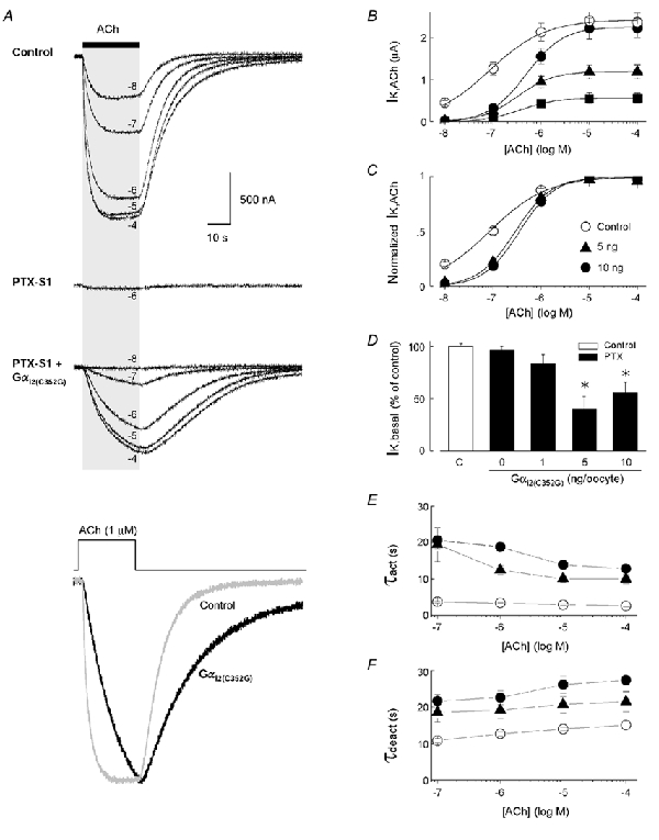
A, typical ACh-evoked GIRK currents (IK,ACh) recorded from different oocytes from three separate experimental groups. Upper traces: IK,ACh from a ‘control’ oocyte expressing muscarinic m2 receptors and Kir3.1/Kir3.2a channel subunits, utilizing endogenous Gαi/o proteins for receptor activation. Middle trace: co-expression of PTX-S1 (1 ng cRNA/oocyte) effectively uncouples ACh-evoked GIRK currents utilizing oocyte Gαi/o proteins. Lower traces: expression of the PTX-insensitive Gαi2(C352G) subunit (5 ng cRNA) with PTX-S1 rescues m2 receptor-coupled GIRK currents. All GIRK currents were elicited by a 25 s application of different concentrations of ACh as indicated. Bottom traces: superimposed IK,ACh elicited by 1 μm ACh from the control oocyte (grey trace) and the Gαi2(C352G)-coupled oocyte (black trace). Peak amplitudes are normalized to illustrate the kinetic differences in the activation and deactivation time courses. B, ACh dose-response relations for GIRK activation via m2 receptors coupled to oocyte Gαi/o subunits (○, control) and Gαi2(C352G) at different levels of expression (▪ 1 ng, ▴ 5 ng, and • 10 ng cRNA/oocyte). C, ACh-dose-response curves from B normalized to maximal IK,ACh. D, comparison of receptor-independent basal GIRK currents (IK,basal) with varying levels of Gαi2(C352G) expression. IK,basal is expressed as the percentage change in the ‘control group’ mean value determined for each batch of oocytes. E, activation time constants (τact) and F, deactivation time constants (τdeact) for GIRK currents coupled to varying levels of Gαi2(C352G) expression and different concentrations of ACh. ○ control; ▴ 5 ng; and • 10 ng of Gαi2(C352G) cRNA/oocyte. Data in B-F represent the means ±s.e.m. from at least 3 batches of oocytes with the number of oocytes indicated. * P < 0.05.