
Figure 3. Biochemical analysis of PTX-insensitive Gαi/o protein levels in Xenopus oocytes.
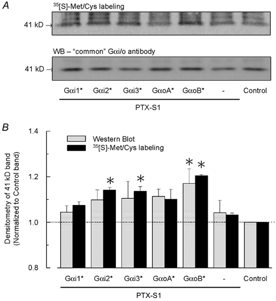
A, radiolabelling (upper panel) and Western blot analysis (lower panel) of oocytes injected with cRNAs encoding five different PTX-insensitive Gαi/o subunits (*) as described in Methods and Fig. 2. Oocytes were incubated for 3 days in OCM containing 0.5 mCi ml−1 [35S]Met/Cys. All groups were injected with cRNAs for the m2 receptor and GIRK channel subunits Kir3.1 and Kir3.2a (0.5 ng each/oocyte). PTX-insensitive Gαi/o cRNAs (5 ng/oocyte) were injected with PTX-S1 cRNA (1 ng/oocyte). Both endogenous and heterologously expressed Gα proteins were immunoprecipitated from the lysate equivalent of one oocyte using a ‘common’ Gα antibody, then separated by SDS-PAGE and transferred to a PVDF membrane for autoradiography and Western blotting. The 41 kDa band corresponding to Gαi/o proteins is indicated. B, quantitative analysis of Gαi/o proteins detected by radiolabelling and Western blot analysis. The 41 kDa band from the control group (no PTX-insensitive Gαi/o cRNA) served as an internal reference and was used to normalize the band intensity among the different experimental groups in each autoradiogram and Western blot. The Western blot results are the means + s.e.m. obtained from 3 independent experiments (separate batches of injected oocytes that were immunoprecipitated and immunostained as described in Methods). The radiolabelling data are the means + s.e.m. obtained from 2 of the Western blot experiments. * P < 0.05.