
Figure 5. External acidification inhibits the inactivation of KCNQ1 current in Xenopus oocytes.
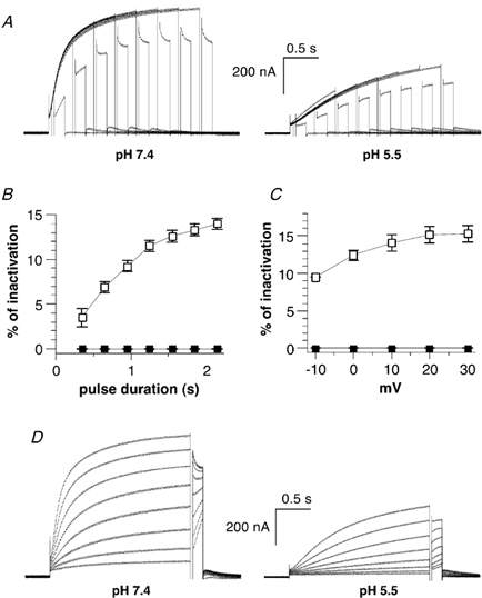
A, inactivation of KCNQ1 was revealed using a three-pulse protocol as in Fig. 4, except that the third test pulse was carried out at -10 mV. The same oocyte was recorded at pH 7.4 (left) and then pH 5.5 (right). B, the percentage of inactivation obtained at pH 7.4 (open squares) and pH 5.5 (filled squares) (n = 8) is plotted as a function of the prepulse duration and calculated as described in Fig. 4C. C, the percentage of inactivation obtained at pH 7.4 (open squares) and pH 5.5 (filled squares; n = 6) is plotted as a function of the prepulse voltage and calculated as described in Fig. 4C. D, the same oocyte was recorded at pH 7.4 (left) and then pH 5.5 (right). A 2 s conditioning prepulse to varying potentials (from -40 to +30 mV, in 10 mV increments) was given, then a brief (20 ms) hyperpolarizing interpulse to -130 mV allowed recovery from inactivation before a test pulse of 150 ms to -10 mV was applied to reopen and reinactivate the channels.