
Figure 2. Location of gigantocellular reticular nucleus (Gi) and dorsal paragigantocellular nucleus (DPGi) sites that produced bilateral muscle atonia, blockage of locomotion and the withdrawal reflex.
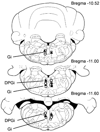
Squares and triangles indicate Gi and DPGi sites that produced bilateral muscle atonia during electrical stimulation (< 100 μA) and microinjections of kainic acid (100 μm, 0.2 μl), respectively. Circles indicate Gi and DPGi sites that suppressed locomotion and the withdrawal reflex.