
Figure 5. Coronal views of the rat midbrain showing the location of recorded RMC and RMC-spinal neurons.
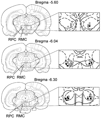
Open and black circles indicate RMC and RMC-spinal neurons, respectively, inhibited by Gi and DPGi stimulation, suppressing locomotion and the withdrawal reflex. Triangles indicate RMC neurons that were excited by Gi and DPGi stimulation. Crosses indicate RMC neurons that were unaffected by Gi and DPGi stimulation.