
Figure 2. S1P activates a pro-inflammatory pathway in RAW 264.7 macrophages.
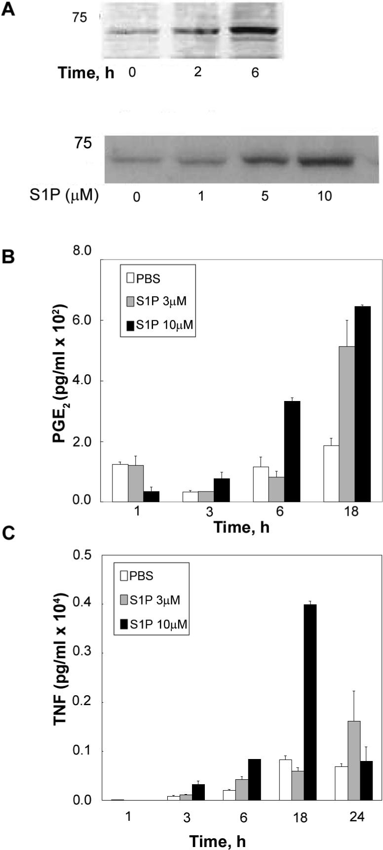
A) Upper panel: RAW 264.7 were treated for 0 to 6h with 1 μM S1P and then the cells were collected and analyzed by western for COX-2 protein levels. Lower panel: RAW 264.7 were incubated for 6.h in the presence or absence of different concentrations of S1P (0−10 μM) and submitted to western blot analysis as described in Experimental Procedures. Results are representative of three independent experiments. B) RAW 264.7 were incubated for up to 18h in the absence or presence of 3 μM or 10 μM S1P. At the end of incubation medium was collected and analyzed for PGE2 levels as described in Experimental Procedures. Results are representative of three independent experiments performed in duplicate. Error bars represent SE. C) RAW 264.7 were incubated for up to 24h in the absence or presence of 3 μM or 10 μM S1P. At the end of incubation medium was collected and analyzed for TNF-α levels as described in Experimental Procedures. Results are representative of three independent experiments performed in duplicate. Error bars represent SE.