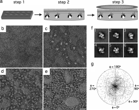
Fig. 1.
Lipid monolayer system for the preparation of single-particle samples. (a) The His-tagged Tf-TfR complex (Tf in black, TfR in white) in aqueous solution is added to a 25-μl well in a Teflon block (step 1). Then, 1 μl of a mixture of Ni-NTA and filler lipids is cast on top of the aqueous solution to form a monolayer (step 2). After incubation at 4°C, an EM grid is placed on the monolayer sample (step 3). The grid is then removed and either negatively stained or frozen in liquid ethane. (b–e) Negatively stained samples of Tf-TfR complex adsorbed to monolayers containing 0% (b), 2% (c), 20% (d), and 40% (e) Ni-NTA lipid. (Scale bar: 30 nm.) (f) Representative class averages of negatively stained Tf-TfR complexes on monolayers. Image 1 shows the predominant top view of the complex. The side length of the images is 27 nm. (g) Angular distribution plot showing that most of the negatively stained His-tagged complexes adopt a preferred orientation on the monolayer.

