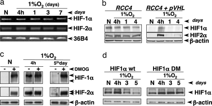
Fig. 2.
Chronic hypoxia promotes HIFα protein degradation depending on the hydroxylation–pVHL–proteasome pathway. (a) Hypoxic kinetics in HeLa cells at 1% O2. HIF-1α, HIF-2α, and 36B4 mRNA levels were analyzed by RT-PCR. (b) RCC4 cells (pVHL-deficient) or the stable clone RCC4-pVHL (which overexpresses pVHL) were incubated in normoxia (20% O2) or hypoxia (1% O2) for different periods of time. (c) HeLa cells were incubated in normoxic (20% O2) or hypoxic conditions (1% O2) for 4 h or 5 days, and 4 h before the lysis, cells were incubated in the presence or absence of DMOG (1 mM). The levels of HIF1α, HIF2α, and β-actin (loading control) were analyzed by Western blotting. (d) 293 cells were incubated in normoxia (20% O2) or hypoxia (1% O2) for different periods of time, and 48 h before the lysis, cells were transfected with the HIF1α WT or DM constructs. The levels of HIF1α were analyzed by Western blotting using the Myc-tag antibody.

