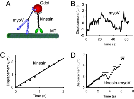
Fig. 1.
Motor movement on microtubules. (A) Cartoon of the experimental setup. Microtubules (MT) are adhered to a glass coverslip. Full-length constructs of myoV, kinesin, or both motors are attached to carboxylated Qdots (see Methods), and visualized by in an objective-type total internal reflectance microscope. Experiments with constitutively active constructs of myoV and kinesin, attached to streptavidin-Qdots via a C-terminal biotin tag, were also performed. (B) One-dimensional diffusion of myoV on microtubules with no net displacement. (C) Smooth movement routinely observed when kinesin alone is attached to the Qdot. Data obtained at the onset of movement when the Qdot appeared on the microtubule until its disappearance when kinesin terminated its run. The velocity for this kinesin molecule was 1.13 μm/s, estimated from the linear regression. Images were captured at 5 frames/s. (D) New behavior seen when both kinesin and myoV are attached to a Qdot (data are from SI Movie 1). For kinesin/myoV-labeled Qdots, numerous runs were characterized by periods of smooth directed movement, as highlighted by the regression through the first 3 s, interspersed with diffusive searches identified by horizontal lines. The periods of smooth movement were characteristic of the movement seen with Qdots labeled only with kinesin (as in C), having velocities (0.80 μm/s for the first 3 s of this run based on regression) comparable to that of kinesin alone (see Table 1). Data obtained at the onset of movement when the Qdot appeared on the microtubule until its disappearance when the run terminated. Note that the scales are different when comparing the Qdot movement here which has both kinesin and myoV attached to that of a Qdot labeled solely with kinesin in C. Image capture rate of 5 frames/s.

