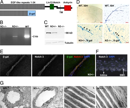
Fig. 1.
Characterization of the Notch 3 knockout mice. (A) A schematic of the heterodimeric Notch 3 receptor (Upper) indicating key structural features. In the extracellular domain, the 34 EGF-like repeats (gray boxes) and the three Lin12-Notch repeats (green boxes) are indicated. The transmembrane domain (TM), and the intracellular ankyrin repeat region are also shown (red boxes). The insertional mutagenesis, which generated the knockout allele, resulted in a fusion protein (Lower) containing EGF-like repeats 1–21 of Notch 3 fused to β-gal (blue box). (B) Long-range PCR amplified intron 16–17 (≈2 kb) of the Notch 3 gene in DNA samples from WT (Notch 3+/+) or heterozygous animals (Notch 3+/−) but failed to amplify the larger intron containing the trapped vector in DNA from knockout mice (Notch 3−/−). (C) The Notch 3 intracellular domain was detected by Western blot analysis of cultured aortic smooth muscle cells (SMCs) derived from WT and Notch 3+/− but absent (also by qRT-PCR, data not shown) in those derived from Notch 3−/− mice. (D) Notch 3 expression. In situ hybridization, using a Notch 3 antisense riboprobe labeled vessels in brain from WT mice (Upper). Likewise, X-gal staining (Lower) of Notch 3+/− (Left) and Notch 3−/− (Right) brain vessels. (E) Immunofluorescence of brain tissue sections demonstrated the colocalization of β-gal and Notch 3 extracellular epitopes in brain arteries from Notch 3+/− mice. (F) Aortic SMC layers from WT mice (white) showed Notch 3 expression. (G) Low magnification electron micrographs of arterial cortical vessels (Left) and aorta (Right) from 8-week-old WT and Notch 3−/− mice. Asterisks indicate smooth muscle cells. L, lumen. (Scale bars: Left, 5 μm; Right, 10 μm.)

