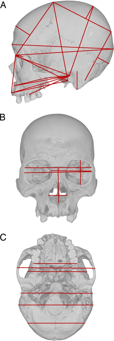
Fig. 1.
The approximate locations of the cranial measurements used in the analyses are superimposed as red lines on lateral (A), anterior (B), and inferior (C) views of a human cranium. Note that when the endpoints of a measurement are not visible, the line is projected into a plane situated in front of the cranium.

