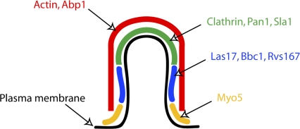
Figure 1.
Schematic model showing the localization of nine proteins on an endocytic invagination. An invagination of intermediate length (∼100 m) is depicted. The coat proteins, including clathrin, coat the tip of the invagination. Rvs167, Las17, and Bbc1 occupy the neck region below the tip. Myo5 concentrates to the base of the invagination. Actin and actin-binding protein Abp1 form a shell covering the whole invagination.

