Figure 1.
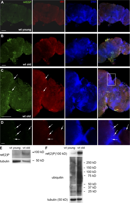
Ref(2)P localization and expression in the adult brain of wild-type flies. (A) Confocal micrographs of adult brain of a young (2 d old) wild-type fly. Positive staining for Ref(2)P and ubiquitin is not evident. (B–D) Confocal micrographs of adult brain of an old (8 wk old) wild-type fly. Ref(2)P accumulates in protein aggregates that often colocalize with ubiquitin (arrows). The white rectangle in C indicates the area shown in D. (E and F) Western blot analysis of total cell lysates (E) and insoluble protein fraction (F) of wild-type young and old adult heads probed with anti-ref(2)P and anti-ubiquitin antibodies, demonstrating a significant increase in Ref(2)P and insoluble ubiquitinated protein levels in old flies. wt, wild type. Bars: (A–C) 100 μm; (D) 10 μm.

