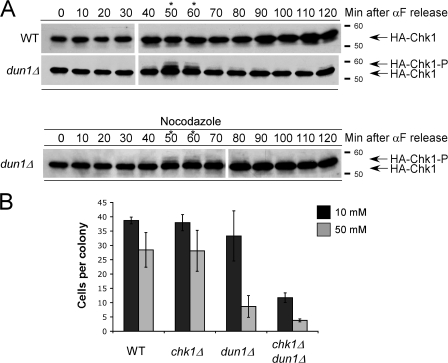
Figure 2.
Chk1p is activated in dun1Δ cells in late S phase and G2 during an unperturbed cell cycle. (A, top) Cells were released from G1. Western blot analysis was performed to monitor HA-Chk1p protein migration. (A, bottom) dun1Δ cells were treated as in the top panel, except cells were released into medium containing nocodazole. The asterisks emphasize the time points at which HA-Chk1p phosphorylation was observed. The Molecular masses (in kilodaltons) are indicated to the right of each panel. (B) Cells were plated on YPD medium containing either 10 or 50 mM HU and incubated at 30°C for 24 h, and the number of cells per colony was counted for 50 colonies. Bars represent the mean number of cells per colony. Error bars represent one standard deviation from the mean.

