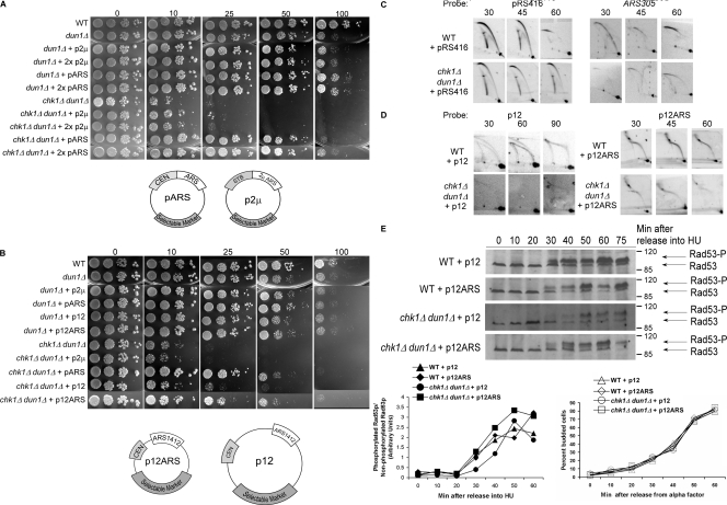
Figure 4.
Suppression of the HU sensitivity of chk1Δ dun1Δ cells by an episomal replication origin activated early in S phase. (A and B) Cells containing the indicated number of copies of pARS (pRS416, pRS415), p2μ (pRS426, pRS425), p12, or p12ARS were spotted onto YPD medium containing the indicated concentration of HU (in millimoles). (C and D) Analyses of replication fork–containing structures were performed as in Fig. 1 B, using probes for the indicated plasmid or ARS305. All cells analyzed in D contained pRS416. (E)Rad53p phosphorylation in response to HU treatment is restored to wild-type levels and kinetics in chk1D dun1D cells by additional replication origins that are activated in early S phase. Rad53p phosphorylation was monitored as in Fig. 3 C. The Molecular masses (in kilodaltons) are indicated to the right of each panel. (E, bottom left) Ratio of phosphorylated to nonphosphorylated Rad53p. (E, bottom right) Budding index.

