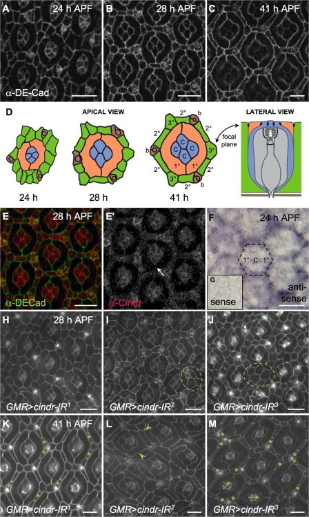
Figure 1.
Patterning the D. melanogaster pupal retina requires Cindr. (A–C and E–G) Wild-type Canton S retinae dissected at the indicated time points. DE-Cad marks AJs. (D) Illustration of ommatidia from A-C showing 1° cells (orange), 2° and 3° cells (green), cone cells (blue), photoreceptors (gray), and bristles (brown). AJs are illustrated in black. Lateral view was inspired by Tepass and Harris (2007). (E) 28-h-APF Canton S retina with Cindr (red) and DE-Cad (green) shown. (E′) Cindr fills the cytoplasm of lattice and cone cells and localizes to AJs; the arrow indicates membrane between two 1° cells. (F) In situ analysis at 24 h APF to detect cindr transcripts; broken line indicates a single ommatidium. (G) Control sense probe. Retinae dissected at 28 (H–J) and 41 h (K–M) APF expressing cindr-IR transgenes as indicated. DE-Cad marks AJs. Mutant phenotypes include failure of IPCs to resolve into single file (circled), excess IPCs (asterisks), discontinuous DE-Cad (arrowheads), and ommatidial misrotation (arrows). Bars 10 μm.

